Begin placing monitors in the following order:
-
Pulse Ox: To monitor O2 saturation, heart rate and peripheral perfusion. A waveform is visible on the anesthesia machine, which can be used to ensure correct placement. The pulse ox is the preferred method to measure heart rate over EKG leads, as the latter may be displaced during surgery or disturbed during cauterization. It measures the ratio of bright red oxygenated hemoglobin to darker red deoxygenated hemoglobin and displays this ratio as percent O2 saturation.
- Location: Same arm as IV.
-
BP Cuff:
- Size = 30% greater than the arm circumference; too large = falsely LOW BP; too small = falsely HIGH BP).
- Location: Opposite arm from IV.
- Senses vibrations ("oscillations") to detect the beginning of turbulent flow/Korotkoff sounds (systolic blood pressure) and the beginning of laminar flow/end of Korotkoff sounds (diastolic blood pressure).
- Measures the Mean Arterial Pressure (when oscillations are greatest), and uses a preset algorithm to calculate the systolic and diastolic blood pressure.
-
5-lead EKG:
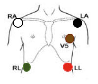
- The EKG creates an electrical field and measures disturbances caused by the heart's electrical activity to create the classic EKG waveform. Classically, V5 is most sensitive to ST changes (as it is directly over the heart), while the lead II is most sensitive to arrhythmias (as the p wave is strongest here).
- A common way to remember lead placement is "Smoke (Black, Brown) over Fire (red)" for the left EKG set, and the opposite color for the right ("White on Right", "Green Opposite Red").
- Place as below:

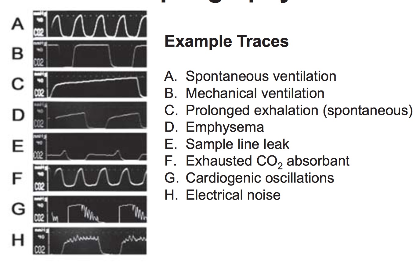
- The fourth monitor is capnography which measures the expired CO2 (i.e. the End Tidal CO2). This is usually connected to the airway circuit (your resident will set this up for you). The ETCO2 provides a waveform which is used to evaluate how well a patient is ventilating. Below is a picture of several ETCO2 waveforms and what they represent. ETCO2 will also fall during periods of low cardiac output as less CO2 is delivered to the lungs.

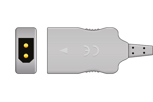
- Other monitors: Temperature probe. Surgery will often place a Foley before positioning the patient after induction; most Foleys have a temperature probe inside the catheter that can be hooked up to the machine. Alternatively, temperature probes can be placed in the axilla, intra-nasally, or in the esophagus. The machine's side of the cord looks like this, except it is usually blue:
